What is magnetic resonance imaging? | Explained Premium
The Hindu
Magnetic resonance imaging is a non-invasive diagnostic tool using magnetic fields to create detailed images of the body's soft tissues.
The story so far: For those trying to look inside the human body without surgery, magnetic resonance imaging is an indispensable tool. The underlying techniques were worked out in the early 1970s; later the same decade, Paul Lauterbur and Peter Mansfield refined them to pave the way for their commercial use. For these efforts, they were awarded the medicine Nobel Prize in 2003, speaking to the significance of the technique and its place in modern medical diagnostics.
Magnetic resonance imaging (MRI) is used to obtain images of soft tissues within the body. Soft tissue is any tissue that hasn’t become harder through calcification. It is a non-invasive diagnostic procedure widely used to image the brain, the cardiovascular system, the spinal cord and joints, various muscles, the liver, arteries, etc.
Its use is particularly important in the observation and treatment of certain cancers, including prostate and rectal cancer, and to track neurological conditions including Alzheimer’s, dementia, epilepsy, and stroke. Researchers have also used MRI scans of changes in blood flow to infer the way the activity of neurons is changing in the brain; in this form, the technique is called functional MRI.
Because of the MRI technique’s use of strong magnetic fields, individuals with embedded metallic objects (like shrapnel) and metallic implants, including pacemakers, may not be able to undergo MRI scans. In fact, if they have a credit card in their pocket, the magnetic fields will wipe its magnetic strip!
An MRI procedure reveals an image of a body part using the hydrogen atoms in that part. A hydrogen atom is simply one proton with one electron around it. These atoms are all spinning, with axes pointing in random directions. Hydrogen atoms are abundant in fat and water, which are present almost throughout the body.
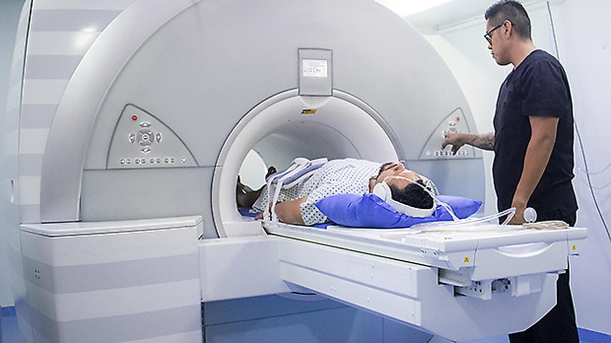
An MRI machine has four essential components. The machine itself looks like a giant donut. The hole in the centre, called the bore, is where the person whose body is to be scanned is inserted. Inside the donut is a powerful superconducting magnet whose job is to produce a powerful and stable magnetic field around the body. Once the body part to be scanned is at the centre of the bore, the magnetic field is switched on.
Each hydrogen atom has a powerful magnetic moment, which means in the presence of a magnetic field, the atom’s spin axis will point along the field’s direction. The superconducting magnet applies a magnetic field down the centre of the machine, such that the axes of roughly half of the hydrogen atoms in the part to be scanned are pointing one way and the other half are pointing the other way. This matching is almost exact: in around a million atoms, only a handful remain unmatched — i.e. a small population of ‘excess’ atoms pointing one way or the other.


 Run 3 Space | Play Space Running Game
 Traffic Jam 3D | Online Racing Game
 Duck Hunt | Play Old Classic Game


